
Hirayama's Disease
Karina Magalhaes de Castro Henriques1, Marco Orsini1,2*, Marcos Raimundo Gomes de Freitas1, Debora Szklarz1, Acary Bulle Oliveira2, Beny Schmidt3, Jano Alves de Souza1, Olivia Gameiro de Souza1, Pedro Moreira1, Camila Pupe1, Osvaldo JN Mascimento1, Eduardo Davidovich1, Carlos Bruno1, Celmir Vilaça1, Amanda Vilas Calheiros1, Bruno Pessoa1, Rossano Fiorelli2, Camila Rodrigues de Almeida2, Pietro Novellino2, Pedro Victor Hugo Bastos4
Affiliation
- 1Department of Neurology, Federal Fluminense University, HUAP, Niterói, Rio de Janeiro, Brazil
- 2Department of Neurology, São Paulo Federal University, UNIFESP, São Paulo, SP, Brazil
- 3Undergraduate of Medicine Federal Fluminense University, Rio de Janeiro, Brazil
- 4Undergraduate of Medicine, Federal Fluminense University, Rio de Janeiro, Brazil
- 5Federal University, Piaiu UFPI, Parnaiba, Brazil
Corresponding Author
Marco Orsini, Masters Program in Science Rehabitation, Department of Neurology, Bonsucesso, Rio de Janeiro, Brazil. E-mail: orsinimarco@hotmail.com
Citation
Orsini, M., et al. Hirayama’s Disease: Case Report ( 2015) Int J Neurol Brain Disord 2(2): 1- 3.
Copy rights
©2015 Orsini, M. This is an Open access article distributed under the terms of Creative Commons Attribution 4.0 International License.
Keywords
lower motor neuron, monomelic amyotrophy, Hirayama disease, spinal muscular atrophy.
Abstract
The Hirayama's disease affects the neurons of the anterior horn of the spinal cord, usually C7 -T1 in young individuals, which causes weakness of the distal muscles of the upper limbs. We report a case of a 14 years old patient presented with significant left hand weakness associated with difficultygripping objects. After performing a resonance of the cervical spine, there were few hyperintense signalsin the anterior horn at the C5-C6 segment. In this situation we can differentiate this case from other conditions, making the correct diagnosis. It is important to report it due to the necessity of better methods that can perform the diagnosis.
Introduction
Hirayama’s disease (HD), also known as monomelicamyotrophy of distal upper limb, is characterized by pure distal motor atrophy of the upper limbs, affecting young men, in the muscles that are innervated by C7, C8 and T1 segments[1-4].It is usually sporadic, it has an insidious onset and there is a slow progression followed by stabilization in 2-4 years. The majority of cases are encountered in Asia, mainly in Japan and India. There are few re¬ports of HD in western countries[1]. The disease is relatively rare and predominantly unilateral, but bilateral cases exists in the literature[2]. In some cases the proximal limb can be affected, more than the distal limb[5-9].
Even though the physiopathology still remains unknown, neuropathologic studies reveal a focal lesion in the anterior horn motor cells of the spinal cord. HD is thought to be caused by a compressive myelopathy, which is developed due to the anterior displacement of the dural sac during the neck’s flexion. Its course is generally benign but a few patients remain disabled[3].
Case Report
KRS, a 14 years old man, student, eight months ago realizes difficulty in some activities that demand dexterity and gripping, just like practicing sports (basketball, swimming) due to muscle weakness. Three months ago, he noticed atrophy of his right forearm and hand (Figure 1). His past medical history was unremarkable with no trauma reference. No family members had neuromuscular complaints. Vital signs were normal during Physical Examination. An atrophy of right hand intrinsic muscles was noticed during Neurological Examination as well as a distal paresis with abolition of stilo radial reflex on the same hand. As the same Examination proceeded, no more abnormalities were registered such as no sensory disorder.
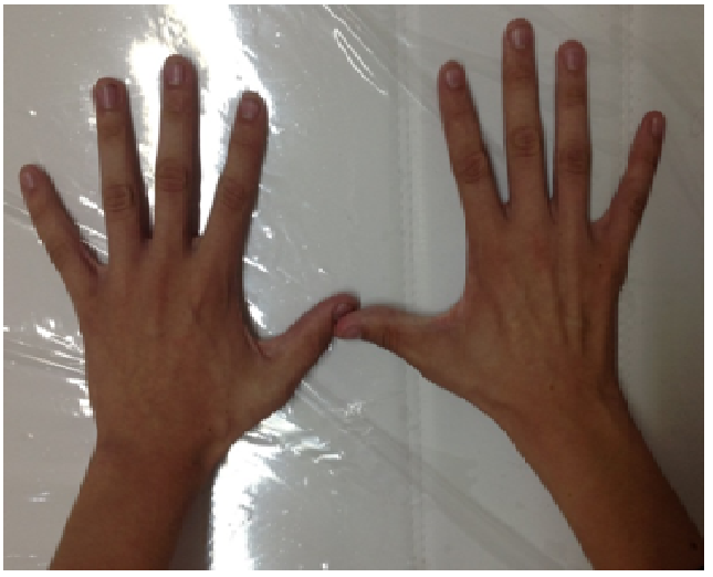
Figure 1: Muscular Atrophy through - right brachial region in patients with 14 years and disease Hirayama
Results
The electromyography showed reduced recruitment, increased amplitude and duration of the motor unit action potentials on the right forearm and hand muscles. We also observed fasciculations in these muscles. The neuroconduction study was normal. Muscle groups of the contralateral upper limb also presented fasciculations potentials. We do not perform the somatosensory evoked potentials study in our patient. The cerebrospinal fluid exam was normal.
The cervical MRI showed a spinal cord bottleneck at the last cervical segments with a T2 signal change (Figure 2). While fleeting the neck, there was a reduction of the dural space.
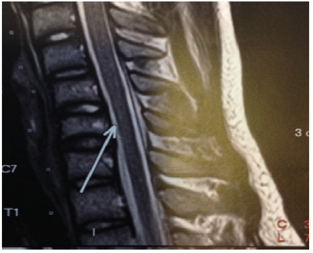
Figure 2: Image cervical spine showing MRI hyper intense region of the anterior horn of the spinal cord at the level of C5-C6
Discussion
Hirayama’s disease was reported for the first time in 1959, but the benign course of the disease allowed larger studies in 1982, with the necropsy findings that showed ischemia of the anterior horn of the spinal cord, neuronal loss and gliosis[5].
Our patient is a 14-year-old male presenting an insidious case of distal atrophy of the upper limb, a typical case ofHirayama’s Disease. The diagnoses of the Benign Monomelic Amyotrophy (HD) were elaborated due to the classic history of paresis and unilateral amyotrophy (one member only). Based on diagnostic criteria, the beginning age associated with the absence of bulbar involvement and/or the pyramidal tract and miotomes of other members allows us to considerate an Amyotrophic Lateral Sclerosis (ALS)[6].
The progressive spinal amyotrophies, because they are symmetric, bilateral and commonly with positive family history, were excluded too. Structural lesions like, for an example, disc protrusions, syringomyelia, cancer and arteriovenous malformations would show sensibility symptoms and also typical images seen at cervical MRI[6].
There are various theories for the disease’s physiopathology, but the mechanism is still uncertain. The most accepted theory is that a fleeting neck biomechanism, acting in both the dural sac and cervical spine, lead us to a circulation disturb and causing lesions at the anterior horn’s cells. This mechanism has been elegantly demonstrated by the study of somatosensory evoked potentials with neck flexion and neutral position. Patients with Hirayama disease have reduced amplitude of the N13 potential during flexion[7]. It’s known that image exams can show parenchyma changes in the low cervical region. Fleeting neck, the findings become more evident with a large reduction of the dural space[8-13]. At the MRI, findings are more evident, with an important reduction of the dural space. Using electrophysiology, we can see a normal sensorial neuro conduction and the motor conduction amplitude may be reduced, specifically the ulnar and median nerves. Besides this studies changes that match with denervation, it is possible to see fibrillation, positives waves and polyphasic potentials[9].
It’s not known why, but HD affects Japanese men. Some authors have related the association with allergy cases, with serum IgE elevation, considering the hypotheses that this disease may be an allergic myelitis. There are few reports of IgA deficiency in patients diagnosed with Hirayama’s disease, but in our patient this dosage was normal[10].
Patients treated with cervical collar, especially those who maintain a flexed neck for long periods of time, have improved. Treatment may still include physical therapy and decompressions might also contribute to clinical improvement[11].
Conclusion
We should be aware that the Hirayama’s Disease or Syndrome mainly affects young male patients who presents distal atrophy of the upper limbs. The patients frequently show improvement with the use of the neck brace, hence it should be introduced as soon as the diagnosis is confirmed.
References
- 1. De Freitas, M.R., Nascimento, O.J. Benign monomelic amyotrophy: a study of twenty-one cases. (2000) Arq neuropsiquiatr 58(3B): 808-813.
- 2. Gamez, J., Pradhan, S. Bilaterally symmetric form of Hirayama disease. (2010) Neurology 74(4): 345.
- 3. Hirayama, K. Juvenile muscular atrophy of distal upper extremity (Hirayama disease). (2000) Intern Med 39(4): 283- 290.
- 4. Hirayama, K., Tsubaki, T., Toyakura, Y., et al. Juvenile muscular atrophy of unilateral upper extremity. (1963) Neurology 13: 373-380.
- 5. Hirayama, K., Toyokura, Y., Tsubaki, T. Juvenile muscular atrophy of unilateral upper extremity: a new clinical entity. Psychiatr Neuro Jpn 61(1959): 2190-2198.
- 6. Hirayama, K., Tomonaga, M., Kitano, K., et al. Focal cervical poliopathy causing juvenile muscular atrophy of distal upper extremity: a pathological study. (1987) J Neurol Neurosurg Psychiatry 50(3): 285-290.
- 7. Restuccia, D., Rubino, M., Valeriani, M., et al.Cervical cord dysfunction during NecK flexion in Hirayama’s disease. (2003) Neurology 60(12): 1980- 1983.
- 8. Ito, S., Kuwabara, T.,Fukutake, Y.,et al. HyperIgEaemia in patients with juvenile muscular atrophy of the distal upper extremity (Hirayama disease). (2005) J Neurol Neurosurg Psychiatry 76(1):132–113.
- 9. Kohno, M., Takahashi, H., Idle, K., et al. Surgical treatment for patients with cervical flexion mielopathy. (1999)J Neurosurg 91(1 Suppl): 33-42.
- 10. Lyu, R.K., Huang, Y.C., Wu, YR.,et al.Electrophysiological features of hirayama disease. (2011) Muscle Nerve 44(2): 185- 190.
- 11. Neves, M.A., Freitas, M.R., Mello, M.P., et al. Benign monomelicamyotrophy with proximal upper limb involvement: case report. (2007)Arq Neuropsiquiatr 65(2B): 524-527.
- 12. Petrova, M., Grigorova, O., Penev, L., et al. Hirayama disease and immunoglobulin A deficiency: a coincidence or a syndrome. (2014) J Neurol Sci 344(1-2): 243- 244.
- 13. Yoshiyama, Y., Tokumaru, Y., Katayama, K., et al. Juvenile muscular atrophy of the bilateral upper limbs associated with peculiar transformation of the dural tube induced by neck flexion. (1994) Rinsho Shinkeigaku 34(1): 65- 71.


