
Oral and Physical Manifestations of a Patient with Trisomy 16: Report of an Unusual Case
Carmen Julia Rovira Ortiz1, Sofia Caraballo Meza2*, Antonio DÃaz Caballero3
Affiliation
1Dentist Periodontology, Department of Periodontology, University of Cartagena, Colombia 2Researcher, Department of Periodontology, University of Cartagena, Colombia
3Doctor of Biomedical Sciences, Department of Periodontology, University of Cartagena, Colombia
Corresponding Author
Sofia Caraballo Meza, Researcher, Department of Periodontology, University of Cartagena, Colombia, USA, E-mail: scaraballom@unicartagena.edu.co
Citation
Caraballo, S.M., et al. Oral and Physical Manifestations of a Patient with Trisomy 16: Report of an Unusual Case. (2016) J Dent & Oral Care 2(2): 1- 4.
Copy rights
© 2016 Caraballo, S.M. This is an Open access article distributed under the terms of Creative Commons Attribution 4.0 International License.
Keywords
Trisomy 16; Partial trisomy 16; Complete trisomy 16; Chromosome; Chromosome 16
Abstract
Clinical report of a 27-year-old patient diagnosed with trisomy 16 and congenital cervical scoliosis; within the oral clinical manifestations of the disease were found agenesis of upper lateral incisors, ankyloglossia, over-inserted labial frenulum and high and arched palate; also the patient showed generalized marginal gingivitis associated to plaque with a loss of attachment level in dental organs 31 and 41.
Partial or complete trisomy 16 are considered non-compatible with life; this anomaly corresponds to 2% of the causes of abortion in the first trimester; SH and DP Roberts Duckett reported a case of survival of 10 months, and in this case, the survival is 27 years.
Introduction
Trisomy is a genetic disorder in which a human being has three homologous chromosomes, instead of a couple that normally exists. Usually one parent has uniparental disomy of the chromosome, in which they have two apparently normal chromosomes but both copies come from only one parent and this results in that there is a trisomy mosaicism in their children, with an extra chromosome in some cells but not in all of them[1,2]. These anomalies are also named genomic mutations, since the number of chromosomes of the genome varies. They can be aneuploidy or polyploidy. The most common case is aneuploidy, that occurs when an individual accidentally presents an extra chromosome (trisomy, 2n+1) or less (monosomy, 2n-1) relative to its normal condition (diploid). The polyploidies occur when the individual has three or more complete sets of chromosomes (Triploidy, 3n; tetraploidy, 4n). In humans, triploidies often end in abortion and if it comes to birth, ends up suffering an early death. Tetraploidy is lethal[3].
Often it has been demonstrated that trisomy 16 is the most frequent in spontaneous abortions surveys. It has been suggested that its lethality is the result of the additional material from the short arm. Trisomy 16 is a genetic anomaly present in 1% of conceptions[2]. The incompatibility with life because of this anomaly makes up approximately 30% of spontaneous abortions due to chromosomal abnormalities, with an estimated incidence in the United States of 100,000 cases annually[4,5].
Based on the literature, very few reliable reports are known about this abnormality and they talk about the visible systemic manifestations in the fetus or the first week of life of the child. Among the anatomical characteristics of the stomatological apparatus diagnosed by some authors are: mouth with thin lips, glossoptosis, arched palate, micrognathia, retrognathia, hypoplastic mandible.
G and Q banding analysis in chromosomes or other cytogenetic banding techniques and also molecular cytogenetics the type of fluorescence in situ hybridization (FISH) and comparative genomic hybridization (CGH), are some of the test or analysis used in the field of genetics including the study of the structure, function and behavior of chromosomes[6].
The test of intellectual functioning: WISC-R, allows an exploration in the verbal area (remote memory deficiencies, understanding and adapting to new social situations, reasoning and numerical calculation, in conceptual relationships, abstract thinking, concentration, understanding and verbal fluency) and non-verbal (memory deficits and visual acuity in capturing causal sequences, in managing spatial relationships and motor skills)[7]. In the clinic case, a trisomy 16 case and its manifestations in the oral cavity and at systemic and cognitive level is reported; for this, a cytogenetic and intellectual functioning study was conducted, which correspond to G and Q banding analysis and the intellectual functioning test: WISC-R, respectively.
Clinic Case
27-year-old male patient turns up with his mother to the Dentistry faculty of the University of Cartagena to the special adult’s clinic; during the anamnesis, the mother refers non-existent consanguinity, or hereditary diseases in the family. She denied to intake mutagens during the course of her pregnancy. She indicated that he is the third child of physically normal parents and born through assisted delivery; at 11 days old he presented a delayed psychomotor development; physical examination presented a size of 1.36 cm and head circumference of 53 cm; macrocranea, flat facie, hypertelorism, antimongoloid palpebral cleft, flat nasal bridge, symmetrical neck, symmetrical thorax, soft abdomen without lumps, micro-penis, testicle in scrotum and normal upper and lower limbs was observed.
An inter consultation was requested due to cognitive and speech deficits; a cytogenetic study was conducted, in this, a peripheral blood sample was taken and cultivated in a RPMI medium, stimulated with phytohemagglutinin and fetal bovine serum and 10% (temperature 37°C); then a G and Q banding was performed, finding a 47 chromosome constitution, XY+ marker chromosome (18, 13, 14, 15 or 16) in the analyzed metaphases; this way, an extra 16 chromosome was identified, with a diagnosis of partial trisomy 16, for which he received a strict medical observation.
At age 8 he was diagnosed with congenital cervical scoliosis at T9-T10 level, the radiographic examination did not show bone malformation, spinal cord displayed shape, position and intensity of signal was normal; spinal dysraphism was not observed; the vertebral canal was noted with dimensions above the usual; the mother reports that the patient did not present medically relevant diseases during his childhood.
At age 21, the WISC-R test was conducted; the verbal area denoted a deficiency in many aspects such as remote memory, understanding and adapting to new social situations, reasoning and numerical calculation, in conceptual relationships, abstract thinking, concentration, comprehension and verbal fluency; in the non-verbal area, he presents deficits in memory and visual acuity, in capturing causal sequences, in managing spatial relationships and motor skills; according to the punctuation obtained with the test, the patient is on average spatial and structure orientation, as well as the associated foresight and motor speed; he presented a moderate cognitive deficit of its intellectual functioning.
Oral Clinical Characteristics
The mother indicated he did not present alterations in the eruption of the temporary dentition; at 6 years old the dental replacement begun, he presented an absence of the upper lateral incisors, during the oral and radiographic examinations it was observed agenesis of the dental organs 12 and 22, over-inserted labial frenum, high and arched palate, secondary caries and a reddened marginal gingiva, with bleeding when stimulated and a generalized enlarged size, except for a decrease in size in the dental organs 31 and 41[Figure 1-3].
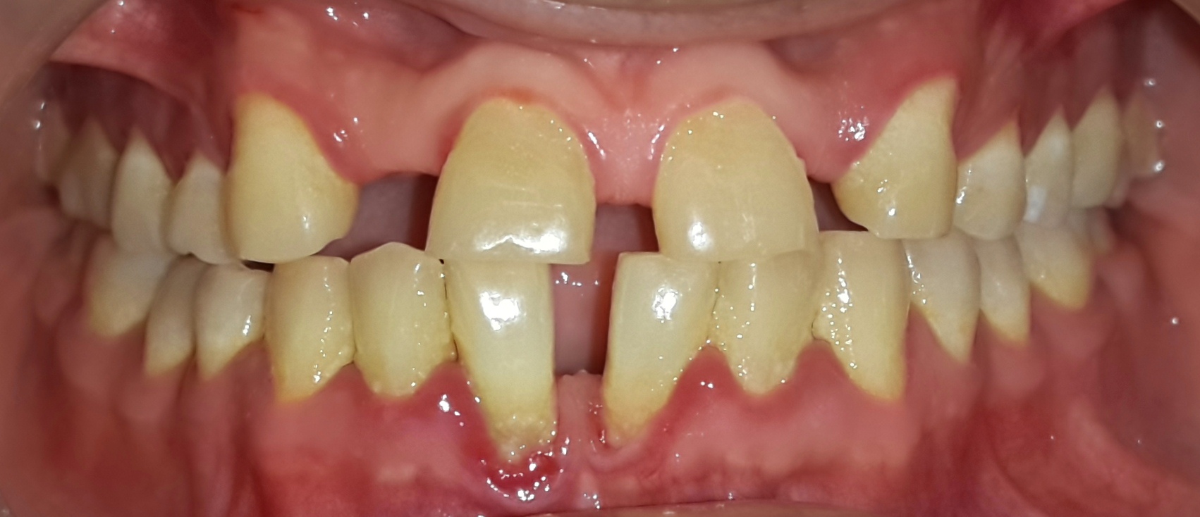
Figure 1: Dental clinical characteristics of the patient.Clinical absence of the upper lateral incisors.
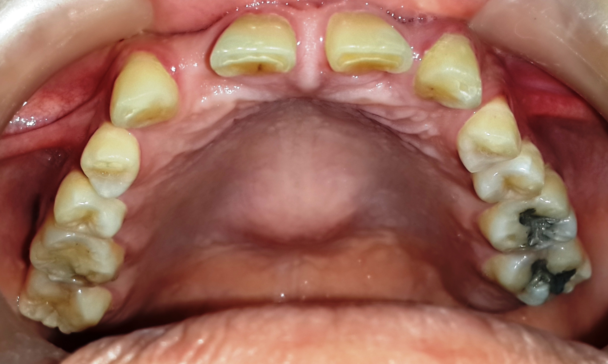
Figure 2: Palate morphology of the patient.Clinical absence of the upper lateral incisors.
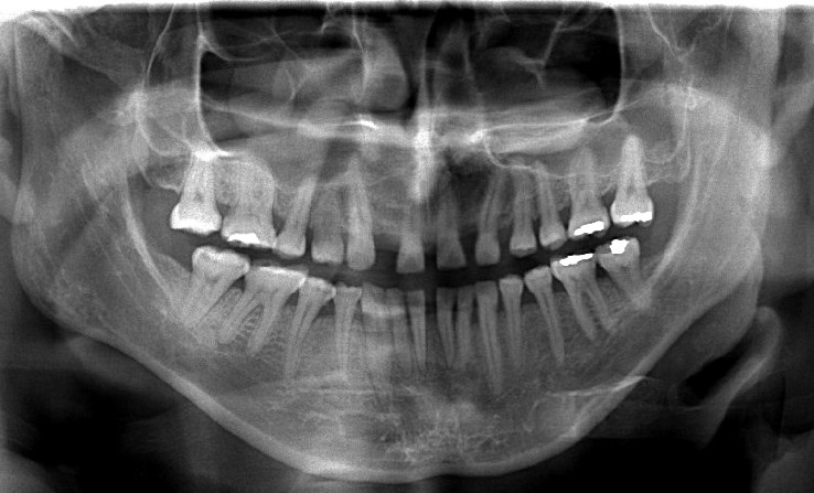
Figure 3: Panoramic radiograph.Agenesis of the upper lateral incisors.
Discussion
In the literature there are many cases of patients with multiple birth defects and developmental delays with partial or complete trisomy 16, only a few of them survive due to systemic conditions and the medical prognosis. In the current clinic case, a patient with agenesis of the upper lateral incisors, ankyloglossia, over-inserted labial ffenulum and high and arched palate was described, these are special stomatological clinical characteristics. Keitaro[8] and collaborators presented a case of a child with trisomy 16, detected by fluorescence in situ hybridization using subtelomeric probes; the patient had abnormal skeletal and facial features, congenital heart defects, imperforate anus, urogenital malformations, delayed postnatal growth and psychomotor retardation.
In addition, he suffered from stenosis of the upper airway due to possible subglottic laryngeal stenosis. The clinical case is developed based on a patient with delayed psychomotor development and congenital cervical scoliosis at T9-T10 level, without any kind of bone malformation.
Lippincott Williams & Wilkins expose the case of an 18-week-old fetus with multiple systemic failures by having complete trisomy 16; at clinical examination, an absent hemi-diaphragm, pulmonary hypoplasia/aplasia, great heart defect, small chest, rib and vertebral defects, cystic kidneys, multiple spleens and imperforate anus were observed together with a cleft palate, nuchal hygroma straps/cystic, microcephaly, marked dysmorphic facial features and big toe dorsiflexion. The detection of partial trisomy 16 denotes special clinical characteristics from the patient, and with that, a different prognosis.
Roberts and Duckett[9] reported the case of an abnormal female baby, who survived for 10 months with an almost complete trisomy 16, at clinical examination of the head and neck, she presented asymmetrical ears, palpebral inclination and a hypoplastic mandible[9]. Finally, the behavior and systemic characteristics, treatment and clinical observation of these type of anomalies allowed a survival of 27 years in the current clinical case.
Conclusion
Partial or complete trisomy 16 is considered non-compatible with life, but in the presented case, the disease behavior and the systemic characteristics changed; the patient was under observation and medical treatment, which influences the increase of survival years. The stomatological characteristics may vary and they are linked to the number of dental organs and the structural morphology of the apparatus.
Acknowledgement:
We are grateful to the University of Cartagena for their cooperation.
References
- 1. Kontomanolis, E.N., Lambropoulou, M., Georgiadis, A., et al. The challenging trisomy 16: a case report. (2012) Clin Exp Obstet Gynecol 39(3): 412-413.
- 2. Been, P. Trisomy 16 and trisomy 16 mosaicism: A review. (1998) Am J Med Genet 79(2): 121-133.
- 3. Chareonsirisuthigu, T., Worawichawong, S., Parinayok, R., et al. Intrauterine Growth Retardation Fetus with Trisomy 16 Mosaicism. (2014) Case Rep Genet.
- 4. Warren, R.J., Rimoin, D.L. The G deletion syndromes. (2012) Journal of Pediatrics 77: 658-663.
- 5. Seller, M.J., Fear, C., Kumar, A., et al. Trisomy 16 in a mid- trimester IVF foetus with multiple abnormalities. (2004) Clin Dysmorphot 13(3): 187-189.
- 6. Stephenson, M., Kutteh, W. Evaluation and management of recurrent early pregnancy loss. (2007) Clin Obstet Gynecol 50(1): 132-145.
- 7. Oliveras-Rentas, R.E., Kenworthy, L., Roberson, R.B., et al. WISC-IV Profile in High-Functioning Autism Spectrum Disorders: Impaired Processing Speed is Associated with Increased Autism Communication Symptoms and Decreased Adaptive Communication Abilities. (2012) Journal of Autism and Developmental Disorders 42(5): 655-664.
- 8. Yamada, K., Uchiyama, A., Arai, M., et al. Severe upper airway stenosis in a boy with partial monosomy 16p13.3pter and partial trisomy 16q22qter. (2009) Congenital Anom(Kyoto) 49(2): 85-88.
- 9. Roberts, S.H., Duckett, D.P. Trisomy 16p in a liveborn infant and a review of partial and full trisomy 16. (1978) J Med Genet 15(5): 375-381.


