
Singleton Pup: A Rare Case of Dystocia in German Shepherd Bitch
Atul Patel1*, Kiran Parmar2, Purushottam Patel1, Jignesh Vadalia3
Affiliation
- 1Department of Veterinary Surgery & Radiology, College of Veterinary Science & A. H., Sardarkrushinagar Dantiwada Agricultural University, Gujarat, India
- 2Veterinary Clinical Complex, College of Veterinary Science & A. H., Junagadh Agricultural University, Gujarat, India
- 3Department of Veterinary Surgery and Radiology, College of Veterinary Science & A. H., Junagadh Agricultural University, Gujarat, India
Corresponding Author
Atul Patel, Assistant Professor, Department of Veterinary Surgery & Radiology, College of Veterinary Science & A. H., Sardarkrushinagar Dantiwada Agricultural University, Deesa-385 535, Gujarat, India, Tel: +91 94083 87407; E-mail: dratulvet07@yahoo.co.in
Citation
Patel, A., et al. Singleton Pup: A Rare Case of Dystocia in German Shepherd Bitch. (2016) J Vet Sci Animal Welf 2(1): 1- 3.
Copy rights
© 2016 Patel, A. This is an Open access article distributed under the terms of Creative Commons Attribution 4.0 International License.
Keywords
Bitch; Caesarean section; Pregnancy; Radiography; Singleton litter
Abstract
This case study describes the dystocia in a German shepherd bitch with a singleton (one puppy) litter with unknown reason. The singleton pup was confirmed by radiography. The complete blood count showed within the normal physiological range. One dead fetus was removed through caesarean section. The aim of this case paper is that pet owner should be visited for normal routine check-up their pet during pregnancy to identify any obvious condition.
Introduction
In general term dystocia means difficult in whelping in bitch, and it may be due to inability to expel fetuses through the birth canal even after full term of pregnancy in bitch[1]. The etiology of dystocia may be maternal or fetal. In bitch dystocia mostly due to uterine inertia that may be partial or complete[2]. The labour ends prematurely in partial uterine inertia while in complete uterine inertia labour period is unable to start. Singleton litters in giant breeds including German Shepherds are rare. This problem is mainly related to the extended duration of the pregnancy and dystocia. Also, it is not easy to predict the time of parturition[3]. A pregnancy is a always need extra care, particularly in a singleton litter, it is considered to be a high-risk pregnancy. A high risk pregnancy may be due to many factors. It may be due to infectious factors, use of advanced age for breeding, history of earlier miss pregnancy, brachycephalic dogs and singleton litters[4,5,6,7]. In the present report, diagnosis and surgical management of dystocia due to singleton pup in German shepherd was described.
Case Description
A three year old, 30 kg, primiparous German shepherd bitch was presented with history of prolonged gestation without labour signs. The bitch was mated before 79 days with a three year old male subsequently after 8th day of heat. On presentation bitch was alert and active. Rectal temperature was 102.6°F. Respiration and heart rates were well within normal physiological limits. Pervaginal examination revealed absence of tonicity in the uterine wall. On 77th days of pregnancy dexamethasone and epidosine was used by local veterinarian. On the next day minute foul smelling greenish discharge started from vagina. Further clinical signs related to parturition were no observed. A ventrodorsal and lateral abdominal radiography was performed to confirm the number and position of fetus. Radiograph revealed single pup in right uterine horn; pup covered whole length of gravid uterine horn (Figure 1). Following diagnosis of singleton pup pregnancy an emergency caesarean section was planned to save the life of bitch before any further major complication occurred.
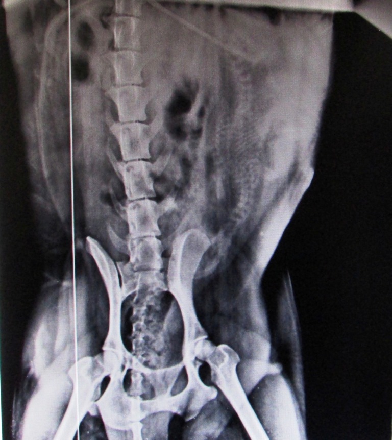
Figure 1: Abdominal radiograph showing single pup within uterus.
Surgical intervention
Preoperatively ceftriaxone(a) (15 mg/kg) and meloxicam(b) (0.5 mg/kg) was given intravenously. The ventral abdomen was shaved and prepared aseptically. As a part of pre- anaesthesia atropine sulphate (0.04 mg/kg) was given intramuscularly, induction was achieved by 5 % isoflurane(C) with help of induction mask and anaesthesia was maintained by 2.5 % isoflurane. Bitch was secured in supine position and caudal mid ventral incision was given. Abdominal cavity was covered with sterile drape to protect any contamination and the gravid uterus was exteriorized (Figure 2). A length of uterine wall incision was given to take out the fetus; unfortunately it was dead and was emphysematous (Figure 3). The uterus was irrigated by normal saline to remove infected uterine exudates material. The inversion patterns of suture in two layers Lambert followed by Cushing were carried out with catgut No. 1/0. The muscles were suture simple interrupted pattern and skin with simple mattress sutures. Post operatively, antibiotic and analgesic treatment was continued for next three days. The skin sutures were removed on postoperative days ten. The case was recovered uneventfully.
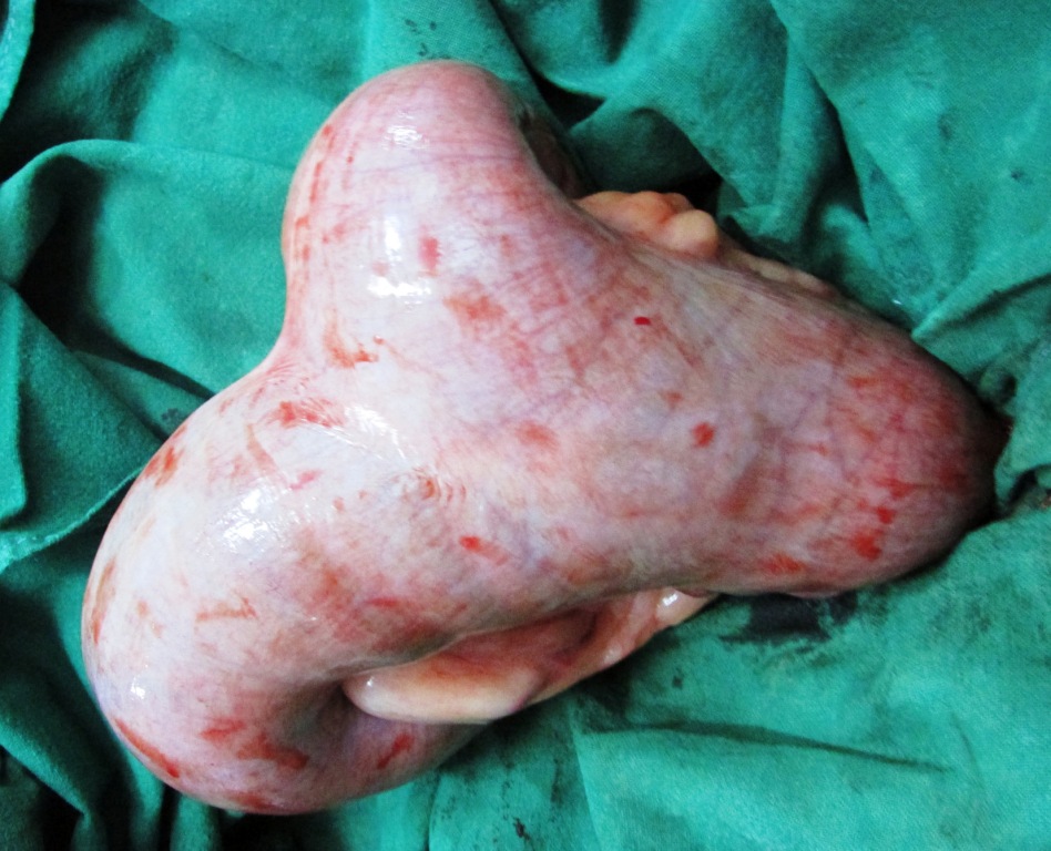
Figure 2: Intraoperative image of exteriorized gravid uterine horn.
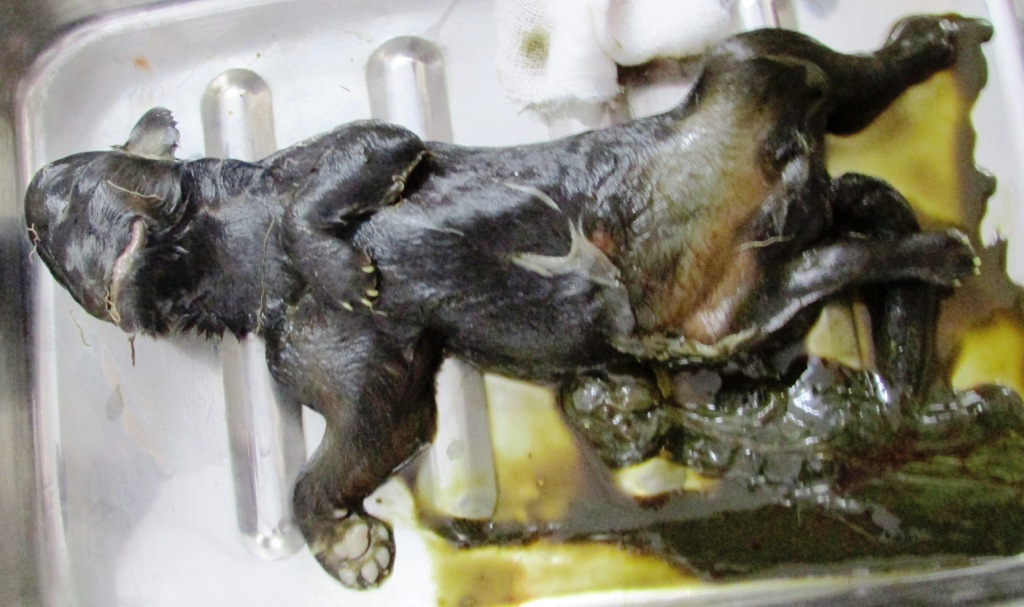
Figure 3: Image of emphysematous pup.
Discussion
In bitches dystocia mostly due to uterine inertia. Two types of uterine inertia; Primary and secondary. In primary uterine inertia a bitch not able to deliver birth to pups after the full term of pregnancy due to the failure of the uterus to begin contraction; in which the uterus fails to start labour in the absence of the fetal signals because of small litter size (single pup syndrome). In single litter the main cause of the dystocia may be overstretching of the myometrium by large size and excessive fetal fluids. The secondary causes of primary uterine inertia may be an inherited predisposition, nutritional imbalance, fatty infiltration of the myometrium, age related changes, deficiency of neuro-endocrine regulation, or systemic diseases in the bitch[8]. In this case radiographic picture shown that a single pup covers the full length of one horn of the uterus. A single litter dystocia due to fails to produce sufficient ACTH and cortisol to initiate the whelping process in bitch. In this case also assumption that not proper production of hormone which play an important role for uterine contraction to start whelping so pregnancy was extended till the caesarean performed. Pup become macerated or mummified due to dies in utero. When infection enters the uterus via the dilated cervix the puppy becomes infected, emphysematous, macerated and only hysterectomy to resolving the case. A greenish vaginal discharge was observed by the pet owner after treatment of local veterinarian; drugs which help to dilation of cervix but due to oversized and emphysematous not able to expelled the pup[9]. A single radiographic exposure is required to diagnose the gravid canine abdomen to detect unlikely harmful to the unborn litter at full term. Radiographically may be detecting number and position of the young ones like fetal maldisposition– such as transverse presentation at the pelvic inlet. Other signs of fetal death, including overlapping of the cranial bones with gas shadows in the fetal heart and stomach and in advanced cases fetal emphysema. In dead fetus the spine is more tightly flexed than the living fetus[9]. Flaccidity of the anterior vaginal wall and failure to stimulate the reflex may indicate presence of uterine inertia. Induction and maintenance with isofurane gave satisfactory result and it was also ideal for such case in which the pup was died. The size of puppy was abnormally large seen in radiography hence site for caesarean section was also important. In this case uterine inertia was related to lack of sufficient quantity of hormonal for initiation of labour pain due to single pup pregnancy and hence caesarean section was performed to relive the condition and save the animal life.
a Intacef, Intas Pharmaceutical, Ahemedabad, Gujarat, India,
b Melonex, Intas Pharmaceutical, Ahemedabad, Gujarat, India
c ISOTROY® 250, Troikaa Pharmaceutical Ltd., Thol, Gujarat, India.
BACK TO TOP↑
References
- 1. Linde Forsberg, C., Eneroth A. Abnormalities in pregnancy, parturition, and the periparturient period. (2000) Textbook of Veterinary Internal Medicine:1527-1538.
- 2. Van den Weijden, B.C., Taverne, M.A.M. Aspects of obstetric care in the dog. (1994) Veterinary Quarterly 16: 20-22.
- 3. Domoslawska, A., Jurczak, A., Janowski, T. A one-foetus pregnancy monitored by Ultrasonography and progesterone blood levels in a German Shepherd bitch: a case report. (2011) Veterinarni Medicina, 56(1): 55-57.
- 4. England, G.C., Russo, M. Ultrasonographic characteristics of early pregnancy failure in bitches. (2006) Theriogenology 66(6-7): 1694-1698.
- 5. Givens, M.D., Marley, M.S. Infectious causes of embryonic and fetal mortality. (2008) Theriogenology 70(3): 270-285.
- 6. Johnson, C.A. High-risk pregnancy and hypoluteoidism in the bitch. (2008) Theriogenology 70(9): 1424-1430.
- 7. Johnson, C.A. Pregnancy management in the bitch. (2008) Theriogenology 70(9): 1412-1417.
- 8. Linde Forsberg, C., Persson, G. A survey of dystocia in the Boxer breed. (2007) Acta Vet Scand 49: 8.
- 9. Peter Jackson, G.G. Dystocia in the dog and cat. (2004) Handbook of Veterinary Obstetrics Second edition 141:166.


